Repair of drooping upper eyelids (ptosis) — both cosmetic and functional correction of levator muscle weakness.
Medically reviewed by EyePlastics Medical Editorial BoardASOPRS oculoplastic surgeonsLast updated June 2026
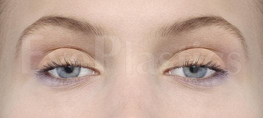
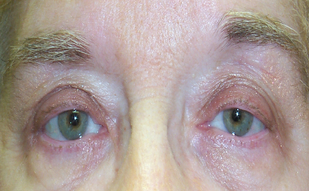
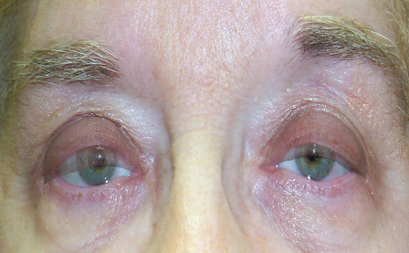
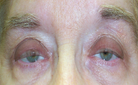
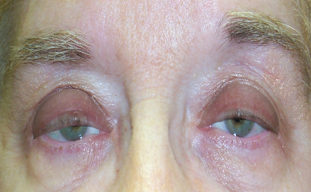
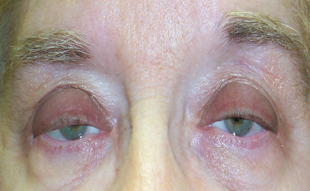
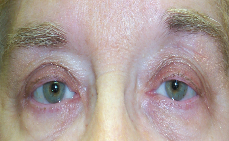
Ptosis (pronounced TOE-sis) is an abnormally low position of the upper eyelid. When the levator muscle — the primary eyelid elevator — or Müller’s muscle weakens, stretches, or detaches from the eyelid, the lid drops in front of the pupil. Ptosis may affect one eye or both and ranges from barely noticeable to severe enough to obstruct vision entirely.
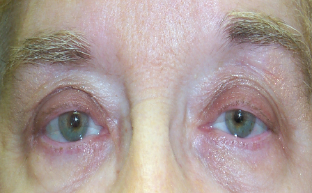
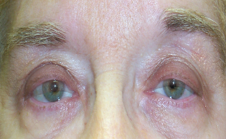
Ptosis is not only a change in appearance — it directly removes vision. Use the simulator below: as you drag the slider, the upper eyelid on the left droops from its normal position into severe ptosis, while the panel on the right shows what the patient actually sees as the lid progressively blocks the upper (superior) field of vision. This superior field loss is what insurers measure with formal visual-field testing when determining whether ptosis repair is functional (covered) rather than cosmetic.
What insurers ask for (functional coverage)
Criteria vary by plan — confirm specifics with your surgeon's office.

Drag the slider — as the eyelid droops, watch how much of the upper visual field disappears.
For a detailed guide to levator anatomy, Müller’s muscle, and the tarsal plate, see our dedicated Anatomy Overview page.
Ptosis is grouped by cause — age-related (aponeurotic), congenital, and neurologic. Recognizing the type guides both the work-up and the operation.
Ptosis ranges from age-related drooping to conditions present from birth, each evaluated and treated differently. Explore each in depth:
Ptosis is more than a cosmetic concern — because the lid sits in the line of sight, it steadily narrows the field of view. Common signs include:
Identifying why the lid droops determines the correct operation, so surgeons classify ptosis by mechanism:
Ptosis (a low lid margin) is distinct from dermatochalasis (excess upper-lid skin), though the two frequently coexist — see Ptosis vs. Blepharoplasty.
A focused eyelid exam measures a few key numbers that drive both the diagnosis and the surgical plan:
Full detail is on our Ptosis Evaluation page.
Treatment is matched to the cause and to levator function. The main options:
See Ptosis Treatment & Surgery for how each is chosen and performed.
Ptosis repair is often functional rather than cosmetic: when a formal visual-field test shows the lid obstructs your superior vision, the repair is frequently covered by insurance with prior authorization and photographs. If ptosis repair is combined with a cosmetic blepharoplasty, the skin (blepharoplasty) portion is billed separately as cosmetic. Ask the office for an itemized estimate and to verify coverage before surgery. The measurements insurers require, self-pay prices, and combined-billing rules are covered in Ptosis Surgery Cost & Insurance, part of our Eyelid Surgery Cost & Insurance guide.
Millimeters decide the result in ptosis surgery, and lid height must be balanced against the fellow eye (Hering’s law can unmask ptosis on the other side after repair). This is best handled by an oculoplastic surgeon with combined ophthalmology and eyelid-plastic training; ASOPRS fellowship-trained surgeons perform these repairs routinely. Find one through our surgeon directory.
Ptosis repair is typically an outpatient procedure under local anesthesia with light sedation. Bruising and swelling settle over 1–3 weeks, and because swelling shifts lid height, the final position is judged at a few weeks to a few months. Most repairs are durable for years; a minority need a minor height adjustment, which is a normal and expected part of fine-tuning millimeter-level symmetry.
The single measurement that drives the surgical plan is levator function — how far the upper lid travels from down-gaze to up-gaze. It sorts patients into three groups, each with a preferred repair:
Two measurements refine it further: MRD-1 (light-reflex to upper-lid margin, normally about 4–5 mm; ptosis at 2 mm or less) sets how much lift is needed, and Hering’s law warns that repairing one lid can unmask ptosis on the other — which an experienced surgeon accounts for before operating rather than discovering afterward. See the full work-up on our Ptosis Evaluation page and the operative detail on Ptosis Treatment.
Connect with a board-certified oculoplastic surgeon who specializes in ptosis.
Search the Directory →