Recognizing and managing complications of the anophthalmic socket — post-enucleation socket syndrome, deep superior sulcus, implant exposure and extrusion, and a poorly fitting prosthesis.
Medically reviewed by EyePlastics Medical Editorial BoardASOPRS oculoplastic surgeonsLast updated June 2026
Part of our complete guide to Anophthalmos & Socket Reconstruction — this page covers anophthalmic socket complications in depth.
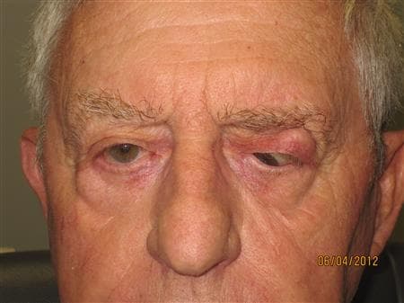
Post-enucleation socket syndrome results from volume loss and structural changes after the globe is removed. The ~7 ml of volume lost is only partially replaced by a standard implant (an 18 mm sphere provides ~3 ml; a 20 mm sphere ~4.1 ml), compounded by up to 3 ml of orbital-fat atrophy over time. Manifestations include:
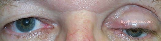
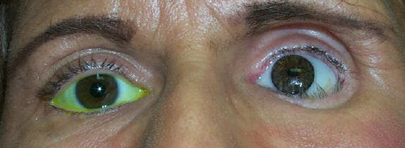
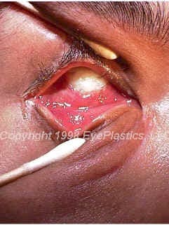
Implant exposure — a breakdown in the conjunctival covering over the implant — is the most common serious complication. A small exposure may be repaired surgically with a patch graft. Larger exposures or progressive extrusion may require implant removal and replacement, often with a dermis-fat graft. Early recognition and treatment improve the chances of preserving the implant.
Connect with a board-certified oculoplastic surgeon who specializes in anophthalmic socket complications.
Search the Directory →