Educational guide to upper facial aging anatomy — brow descent, eyelid hooding, tear troughs, and temple hollowing — and treatments that restore youth.
Medically reviewed by EyePlastics Medical Editorial BoardASOPRS oculoplastic surgeonsLast updated June 2026
The aging eye is rarely about the eye itself. When patients say they look “tired,” “angry,” or “sad” despite feeling well-rested, they are usually describing a constellation of predictable anatomic changes in the upper face. Understanding why the periocular region ages the way it does is the first step toward choosing treatment that actually addresses the cause — rather than chasing a surface symptom.
Upper facial aging is the product of four parallel processes occurring simultaneously: gravitational descent of soft tissue, volume loss in fat compartments and bone, skin changes (loss of elastin and collagen, sun damage), and repetitive muscle activity that etches dynamic lines into static ones. These four forces act on a complex three-dimensional scaffold of bone, retaining ligaments, fat pads, muscle, and skin. Treating only one layer — for example, lasering the skin while ignoring underlying volume loss — produces results that look incomplete or even unnatural.
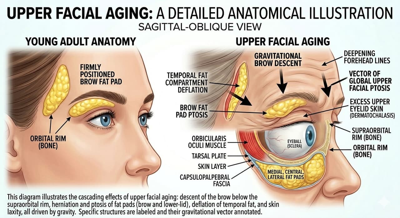
Oculoplastic surgeons evaluate the upper face from top to bottom, layer by layer, treating each finding in the context of the whole. The sections below walk through this anatomic sequence — from brow to temple — and then map each change to a treatment ladder ranging from injectables to surgery.
The brow is the upper frame of the eye, and its position dictates the apparent fullness of the upper lid. In youth, the female brow typically sits at or just above the superior orbital rim with a gentle lateral arch; the male brow sits flatter and lower, directly on the rim. With age, the forehead soft tissues lose ligamentous support and descend, pulled inferiorly by gravity and the depressor muscles (corrugator, procerus, and orbicularis oculi).
The lateral brow descends first and most noticeably because it lacks the support of the frontalis muscle, which inserts more medially. This lateral hooding overhangs the temporal eyelid and creates a heavy, fatigued appearance. Many patients unconsciously elevate the frontalis chronically to lift the brow out of their visual field — this is the origin of deep horizontal forehead lines.
Treating the upper lid without addressing brow position can produce a hollowed, surprised look. Learn more about Brow Lift procedures and how brow position influences blepharoplasty planning.
Upper eyelid hooding is the redundant fold of skin and orbicularis muscle that drapes over the eyelid crease and, in advanced cases, the lashes themselves. It is caused by a combination of dermatochalasis (excess eyelid skin), descent of the brow contributing additional tissue from above, and forward herniation of the orbital fat pads through a weakened orbital septum.
True Ptosis — drooping of the eyelid margin itself due to levator muscle disinsertion — is a distinct entity from hooding and is present in roughly 20–30% of patients seeking blepharoplasty. Missing co-existing ptosis is one of the most common reasons for a disappointing cosmetic result. An oculoplastic surgeon measures margin-to-reflex distance (MRD1), levator function, and lid crease position to differentiate these conditions.
The upper lid has two fat compartments: the medial (nasal) pad, which is characteristically paler/whiter, and the central (preaponeurotic) pad, which is more yellow. The lateral “fullness” over the lateral lid is not fat — it is descended brow tissue and lacrimal gland prolapse, and must be managed differently.
The lower eyelid undergoes some of the most visually impactful changes of the aging face. Three fat pads — medial, central, and lateral — sit behind the orbital septum. With age, the septum weakens and the fat herniates forward, producing the “bags” under the eyes. Simultaneously, the cheek descends, exposing the orbital rim and creating a sharp transition (the “lid-cheek junction”) between bulging fat above and hollowed cheek below.
The skin of the lower lid is the thinnest on the body — about 0.5 mm — and shows fine wrinkling (rhytids), pigmentation changes, and festoons (chronic edema of the malar skin) earlier than other areas. Hyperpigmentation, often misdiagnosed as “dark circles,” may be due to thin skin revealing underlying orbicularis muscle, vascular congestion, or a true tear trough shadow.
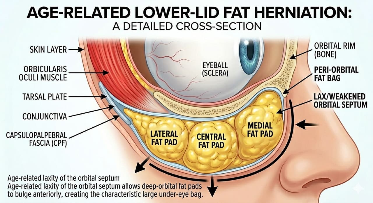
The tear trough is the depression along the inferomedial orbital rim, running from the medial canthus diagonally toward the cheek. It is created by the firm attachment of the orbicularis retaining ligament to the bony rim, which tethers the skin while surrounding tissues descend or herniate. In youth, a robust medial cheek fat pad camouflages this transition; with age, that fat deflates and the trough becomes a visible groove.
Patients often describe the tear trough as “dark circles,” but the darkness is actually a shadow cast by overhanging fat above and hollowness below — not a pigmentation problem. This is why concealer and brightening creams rarely help. Restoring volume across the trough, either with Fillers or surgical fat repositioning, eliminates the shadow at its source.
Important: Tear trough filler is one of the most technique-sensitive injections in cosmetic medicine. Overfilling, superficial placement, or use of hydrophilic products causes the dreaded blue-gray “Tyndall effect” or persistent puffiness that can last years. This area should be treated by injectors with deep periocular anatomic expertise.
The midface — the region between the lower lid and the upper lip — is anchored by retaining ligaments that weaken with time. As these ligaments stretch, the malar fat pad slides inferiorly and medially, deepening the nasolabial fold, hollowing the upper cheek, and unmasking the inferior orbital rim. The result is the classic “double-convexity” deformity: bulging lower lid fat above, a depressed lid-cheek junction in the middle, and a sagging cheek below.
This is why patients in their 50s and beyond rarely look refreshed after lower blepharoplasty alone — the bag is gone, but the underlying skeletal hollowness is now more visible. Addressing midface descent often requires either substantial volume restoration with filler or fat grafting, or a formal midface lift performed through a transconjunctival or temporal approach.
The temples are an often-overlooked component of upper facial aging. The temporal fossa contains a small fat pad and the temporalis muscle; both atrophy with age, while overlying skin and superficial fat also deflate. The result is a hollow, “skeletonized” appearance lateral to the brow, which causes the lateral brow to drop further (loss of its underlying support) and emphasizes crow’s feet.
Restoring temple volume with deep filler placement is one of the highest-impact facial rejuvenation interventions available, though the temple contains significant vessels and requires careful technique by an experienced injector to minimize vascular risk. A modest amount of product at the deep temporal fascia plane lifts the lateral brow, softens the transition from forehead to cheek, and dramatically reduces the “ill” or gaunt look that hollow temples create.
Once the anatomic diagnosis is complete, treatment follows a logical ladder from least to most invasive. The right rung depends on the patient’s age, severity of findings, downtime tolerance, budget, and goals. Most patients benefit from a combination across multiple rungs over time.
| Anatomic Change | Non-Surgical | Minimally Invasive | Surgical |
|---|---|---|---|
| Brow descent | Botox to depressors | Thread lift, RF tightening | Brow lift (endoscopic, direct, temporal) |
| Upper lid hooding | — | Plasma skin tightening | Upper blepharoplasty ± ptosis repair |
| Lower lid bags | Camouflage filler at rim | Laser resurfacing | Transconjunctival lower blepharoplasty |
| Tear trough | HA filler | — | Fat repositioning blepharoplasty |
| Midface descent | Cheek filler | Threads, ultrasound (Ultherapy) | Midface lift, SOOF lift |
| Temple hollowing | Deep temple filler | Fat grafting | Temporal lift |
Neuromodulators (botulinum toxin) are the cornerstone of non-surgical upper facial rejuvenation. Strategic weakening of the corrugator, procerus, and lateral orbicularis allows the frontalis to elevate the brow unopposed — producing a subtle “chemical brow lift” of 1–3 mm at the lateral brow. Botox also softens glabellar “11” lines, forehead lines, and crow’s feet. Learn more about Botulinum Toxin applications.
Hyaluronic acid fillers address volume loss. Different products are designed for different depths and tissues: firm, structured products (Restylane Lyft, Voluma) belong on bone in the temple and midface; soft, low-G-prime products (Restylane-L, Volbella, Restylane Eyelight) are appropriate for the tear trough.
Skin treatments — medical-grade retinoids, sunscreen, vitamin C, and chemical peels — address pigmentation and fine lines but cannot reverse volume loss or descent. They are best understood as foundational maintenance, not transformative.
Between injectables and surgery sits a growing category of energy-based and threaded interventions.
These technologies are excellent adjuncts but rarely replace surgery in patients with significant tissue redundancy or fat herniation. Expect 10–30% of the result a comparable surgical procedure would deliver.
Blepharoplasty remains the gold standard for definitive correction of eyelid hooding and lower lid fat herniation. Upper blepharoplasty removes excess skin and conservative amounts of fat through a hidden incision in the lid crease; lower blepharoplasty is most often performed transconjunctivally (from inside the lid) to avoid an external scar and preserve lid margin position. Modern technique emphasizes fat repositioning over removal, redraping herniated fat into the tear trough to restore a smooth lid-cheek junction.
Brow lift techniques range from direct excision above the brow (best for heavy male brows or asymmetry), to temporal lift through a hairline incision (ideal for isolated lateral descent), to endoscopic lift with small scalp incisions (full forehead). Choice depends on hairline position, forehead height, and severity of descent.
Midface lift elevates the descended malar fat pad and SOOF (sub-orbicularis oculi fat) to recreate youthful cheek projection and close the lid-cheek junction. Performed through a transconjunctival or temporal approach, it pairs naturally with lower blepharoplasty in patients with combined deformity.
Patients with significant Eyelid Laxity often require canthal tightening at the time of lower blepharoplasty to prevent post-operative lid malposition. A thorough pre-op exam identifies this need.
The best treatment plan begins with an honest assessment of three variables: what bothers you, what the anatomy actually shows, and how much downtime is acceptable. A patient in her 40s with early tear trough shadowing and mild brow descent may achieve excellent results with Botox and a single syringe of filler. A patient in her 60s with deep hooding, fat herniation, and midface descent will not be served by injectables alone — she needs surgery, and trying to filler her way out of the problem leads to the overfilled, “puffy” look that has become a cautionary tale in modern aesthetic medicine.
Equally important is sequencing. When surgery and injectables are both indicated, surgery generally comes first — addressing structural problems — followed by fine-tuning with volume and skin treatments. Operating around large volumes of filler can be challenging, and filler in surgical fields can be displaced or partially metabolized by the procedure.
The single most important decision is choosing the right surgeon. The eyelids and periocular region are unforgiving; millimeters matter, and the functional consequences of a poor outcome — dry eye, lagophthalmos, ectropion, asymmetric brow — can be permanent. An ASOPRS fellowship-trained oculoplastic surgeon brings dedicated subspecialty expertise in the anatomy and function of this region.
Whether you are exploring your first injectable treatment, considering blepharoplasty, or seeking a second opinion on a more complex plan, a thorough consultation with a qualified specialist is the essential first step. Find a Doctor in your area to discuss your goals and develop a personalized roadmap for upper facial rejuvenation.
Connect with a board-certified oculoplastic surgeon who specializes in upper facial aging.
Search the Directory →