Infections of the eyelid (preseptal) and orbit (orbital cellulitis) — how they differ, why orbital cellulitis is an emergency, and how each is treated.
Medically reviewed by EyePlastics Medical Editorial BoardASOPRS oculoplastic surgeonsLast updated June 2026
Part of our complete guide to Eyelid & Orbital Infections — this page covers preseptal and orbital cellulitis in depth.
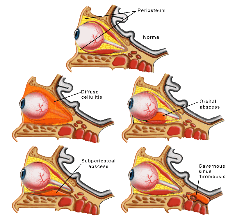
Infections around the eye are classified by one anatomic landmark: the orbital septum — a fibrous sheet that runs from the orbital rim into the eyelids and walls off the eyelid soft tissue from the orbit itself. Infection in front of the septum (preseptal cellulitis) is usually managed with antibiotics alone. Infection behind the septum (orbital cellulitis) surrounds the eye, the extraocular muscles, and the optic nerve — and is treated as an emergency.
Preseptal cellulitis is an infection of the eyelid and periorbital soft tissue. It typically follows a skin source — an insect bite, stye (hordeolum), chalazion, trauma, or spread from the lacrimal sac (dacryocystitis) or sinuses. Common organisms include Staphylococcus aureus, Streptococcus pneumoniae, and (less often since routine immunization) Haemophilus influenzae.

Older children and adults with mild preseptal infection are treated with oral antibiotics and close follow-up; patients should seek immediate care if they develop pain with eye movement, double or decreased vision, bulging of the eye, or fail to improve within 24–48 hours, as these may signal progression to orbital cellulitis. Infants, the unimmunized, and any patient who worsens on oral therapy are admitted for intravenous antibiotics.
Orbital cellulitis is infection of the tissues behind the septum. Roughly 90% of cases extend from bacterial sinusitis (especially the ethmoid sinuses, separated from the orbit by paper-thin bone); the remainder follow trauma, surgery, or spread from adjacent infection.

Any suspicion of orbital involvement warrants CT of the orbits and sinuses — to confirm sinus disease, look for a subperiosteal or orbital abscess that would need surgical drainage, exclude a retained foreign body after trauma, and rule out a mass. Blood cultures are drawn before antibiotics. Vision, color vision, pupils, eye movements, and intraocular pressure are monitored serially — deterioration despite IV antibiotics suggests abscess formation.
An oculoplastic surgeon manages the orbital side of these infections — monitoring the optic nerve, draining abscesses, and coordinating care with ENT and infectious disease.
Connect with a board-certified oculoplastic surgeon who specializes in preseptal & orbital cellulitis.
Search the Directory →