Infections of the tear-drainage system (dacryocystitis, canaliculitis) and traumatic injuries such as canalicular lacerations, and how they are repaired.
Medically reviewed by EyePlastics Medical Editorial Board·ASOPRS oculoplastic surgeons·Last updated June 2026
Part of our complete guide to Tear-Duct & Lacrimal Surgery — this page covers lacrimal infections and trauma in depth.
Infections of the Lacrimal System
Dacryoadenitis (Lacrimal Gland Infection)
Inflammation or infection of the lacrimal gland in the superolateral orbit
Acute: bacterial (Staphylococcus, Streptococcus) or viral (EBV, mumps, CMV, VZV); painful S-shaped lid deformity; treat with systemic antibiotics or antivirals
Chronic: associated with systemic disease — sarcoidosis, Sjögren’s syndrome, lymphoma, IgG4-related disease; biopsy of the lacrimal gland is often required to establish diagnosis
Canaliculitis
Canaliculitis is infection of the canaliculus (the channel connecting the punctum to the lacrimal sac). It is commonly misdiagnosed and undertreated — always consider it when a patient has a “chronic conjunctivitis” of one eye that fails to respond to drops.
Most common cause:Actinomyces israelii is the single most commonly identified organism, forming the characteristic sulphur-granule concretions within the canaliculus; Staphylococcus, Streptococcus, and mixed flora are also frequent
Other causes: Propionibacterium, Fusobacterium, Candida, Aspergillus, herpes simplex
Classic signs: unilateral red eye, mucopurulent discharge, swollen pouting punctum (the “lacrimal pouch sign”); yellow concretions expressed from the punctum on compression
Diagnosis: clinical — probe passes easily but with a “gritty” sensation; microscopy and culture of expressed material confirms organism
Treatment: canaliculotomy (incision of the canaliculus through its posterior wall) with curettage of concretions, followed by irrigation with penicillin or povidone-iodine. Topical antibiotics alone almost always fail. Incomplete concretion removal leads to recurrence
Dacryocystitis — Acute
Acute dacryocystitis is a bacterial infection of the lacrimal sac, almost always arising from nasolacrimal duct obstruction with stasis of tears and secondary infection.
Presentation: sudden-onset pain, redness, and tender swelling at the medial canthus below the medial canthal tendon (this location distinguishes dacryocystitis from ethmoid sinusitis or subcutaneous abscess, which present above or along the tendon)
Common organisms: Staphylococcus aureus (most common), Streptococcus pneumoniae, Haemophilus influenzae, gram-negative rods in immunocompromised patients
Treatment:
Oral antibiotics (Augmentin, Keflex) for mild-to-moderate cases
IV antibiotics (nafcillin, vancomycin for MRSA coverage) for severe disease, periorbital spread, or failure of oral therapy
Warm compresses
Do not probe an acutely infected system — probing risks spreading infection and creating a fistula
Incision and drainage if abscess forms and is fluctuant
DCR surgery after the acute infection resolves (typically 4–6 weeks later) to prevent recurrence
Complications if untreated:
Preseptal (periorbital) cellulitis
Orbital cellulitis and abscess (sight- and life-threatening)
Lacrimal fistula (spontaneous drainage through the skin)
Mucocele (chronic distended, obstructed sac without acute infection)
Cavernous sinus thrombosis (rare but potentially fatal)
Dacryocystitis — Chronic
A chronically obstructed lacrimal sac that is distended with mucoid or mucopurulent fluid with minimal acute inflammation
Presents as recurrent episodes of mild discharge and epiphora, with a soft, compressible swelling at the medial canthus
Pressure on the sac expresses mucoid material through the punctum (regurgitation test positive)
May harbor dacryoliths (stones) from Actinomyces or Candida species
Treatment: DCR surgery; topical antibiotics provide only temporary symptomatic relief
Lacrimal Trauma — Canalicular Lacerations
Canalicular lacerations occur when trauma to the medial eyelid severs the canaliculus. Because the canaliculus lies just beneath the skin medial to the punctum, even seemingly superficial medial eyelid lacerations frequently involve it. If not repaired promptly and correctly, permanent epiphora results.
Recognition
Any laceration medial to the punctum should be assumed to involve the canaliculus until proven otherwise
Common mechanisms: dog bites (very high frequency of canalicular involvement), fist injury, motor vehicle accident, fishhook
The lower canaliculus is injured more commonly than the upper
Both canaliculi may be involved if trauma crosses both lids
Repair Principles
Canalicular repair must be performed within 24–48 hours for best results. The key steps:
Identify both ends of the lacerated canaliculus under magnification (surgical microscope or loupes)
Place a stent (silicone tube) to maintain the lumen during healing and prevent stricture
Reapproximate the pericanalicular tissue (and, when feasible, the canalicular ends) over the stent with fine sutures
Repair the eyelid in layers
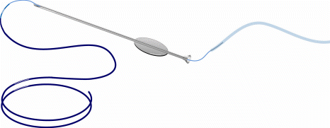
Stent Options
FCI Ophthalmic self-stable (‘autostable’) bicanalicular intubation set used for canalicular and lacrimal intubation.
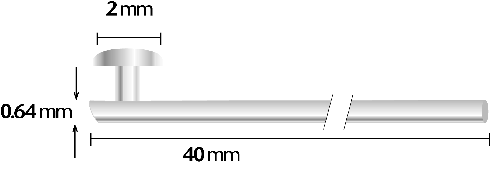
Monocanalicular stent (Mini-Monoka): stent placed only in the injured canaliculus; plugs at the punctum; avoids potential damage to the normal canaliculus. Preferred for isolated lower canalicular laceration
Bicanalicular stent: looped through both upper and lower canaliculi; retrieved nasally. Required when the common canaliculus or lacrimal sac is involved. Disadvantage: potential trauma to the uninvolved canaliculus
Stents removed at 3–6 months
Success Rate
Prompt, meticulous primary repair achieves functional patency in approximately 85–90% of cases
Delayed repair or failure of initial repair may require DCR or ultimately Jones tube if the canaliculus cannot be reconstructed
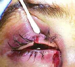
Canalicular Laceration Repair — Clinical Sequence
Eyelid lacerations near the inner corner frequently cut the canaliculus — the tear-drainage channel. Repair requires microsurgical reapproximation over a soft silicone stent, which keeps the channel open while it heals.
Laceration involving the canaliculusThe cut canalicular ends are identifiedA silicone stent is threaded through the system
The lid is repaired over the stentStent introduction (diagram)Final stent position (diagram)
Frequently Asked Questions
What is dacryocystitis?
An infection of the tear sac, usually behind a blocked tear duct — causing pain, redness, and swelling at the inner corner of the eye. It is treated with antibiotics and, once settled, often DCR surgery to remove the underlying blockage.
What happens if a tear-duct (canalicular) laceration isn't repaired?
A canalicular laceration that is not repaired promptly can heal with a permanent blockage and chronic tearing. Timely microsurgical repair over a silicone stent restores the drainage channel.
Find a Specialist
Connect with a board-certified oculoplastic surgeon who specializes in tear-sac infections & lacrimal trauma.