Non-cancerous eyelid growths — including xanthelasma and molluscum contagiosum — and how they are evaluated and removed.
Medically reviewed by EyePlastics Medical Editorial BoardASOPRS oculoplastic surgeonsLast updated June 2026
Part of our complete guide to Eyelid Skin Tumors — this page covers benign eyelid lesions in depth.
Most eyelid lesions are benign. They are grouped by tissue of origin — epithelial, glandular, vascular, and inflammatory. The key clinical task is distinguishing benign from pre-malignant or malignant lesions, which can look deceptively similar.
Squamous papilloma (skin tag) is the most common benign eyelid lesion — a soft, pedunculated or sessile growth arising from the epidermis, often at the lid margin or medial canthus. Some are associated with HPV, though many are not; it is treated by simple snip excision.
Seborrheic keratosis appears as a waxy, “stuck-on” brown plaque with a velvety or verrucous surface. It is benign and may be observed; excision or cryotherapy is performed if symptomatic or cosmetically bothersome.
Keratoacanthoma is a rapidly growing, cup-shaped nodule with a central keratin plug that appears over weeks, then may involute. Because it can be clinically indistinguishable from squamous cell carcinoma, excision with pathologic confirmation is required.
Actinic keratosis is a pre-malignant epithelial lesion caused by cumulative UV damage. It appears as a rough, erythematous, scaly patch on sun-exposed skin. Untreated, a small percentage progress to squamous cell carcinoma over time. Treatment options include cryotherapy, topical field therapies (e.g., 5-fluorouracil, imiquimod, photodynamic therapy), and excision when malignancy cannot be excluded.
Epidermal inclusion cysts are smooth, firm, skin-colored nodules formed by trapped keratinizing epithelium. They are the most common eyelid cysts and are treated by complete excision of the cyst wall.
Dermoid cysts are congenital choristomas containing hair follicles and adnexal structures. They classically occur at the supero-temporal orbital rim (frontozygomatic suture) or supero-medially. Dermoids do not involute and are excised electively — rupture causes severe granulomatous inflammation.
Sweat ductal cysts (eccrine or apocrine hidrocystomas) appear as translucent, fluid-filled cysts along the lid margin. They may enlarge in warm weather. Marsupialization or excision is curative.
Capillary hemangioma (infantile hemangioma) is the most common orbital and periorbital tumor of childhood. It proliferates rapidly in the first year of life, then slowly involutes — about 90–95% resolved by age 9. If the lesion causes amblyopia, astigmatism, or significant ptosis, early treatment with oral propranolol is indicated.
Port-wine stain (nevus flammeus) is a vascular malformation present at birth and does not involute. Extensive port-wine stains in the V1 distribution may be associated with Sturge-Weber syndrome and require glaucoma screening. Pulsed-dye laser is the primary treatment.
Pyogenic granuloma appears as a rapidly growing, red, bleeding vascular nodule, typically after minor trauma or surgery. Despite the name, it is not infectious. Treatment is excision.
Eyelid nevi may be junctional, compound, or intradermal. A lid margin nevus is common and often stable for years. Indications for excision include change in size, shape, color, or bleeding — all of which raise concern for melanoma. The nevus of Ota (oculodermal melanocytosis) involves periocular skin and the episclera and carries a small risk of uveal melanoma; ophthalmologic follow-up is recommended.
Xanthelasma palpebrarum are yellowish, soft plaques of lipid-laden macrophages (foam cells) deposited in the superficial dermis of the medial eyelids. They are the most common form of cutaneous xanthoma. Approximately 50% of patients with xanthelasma have an underlying lipid disorder (hypercholesterolemia, hypertriglyceridemia, or mixed dyslipidemia) — fasting lipid panel and cardiovascular risk assessment are appropriate at initial evaluation.
Xanthelasma is benign and carries no risk of malignant transformation. Indications for treatment are cosmetic. Options include:
Recurrence is common regardless of treatment modality, especially if the underlying dyslipidemia is not controlled. Statin therapy or dietary modification may slow recurrence but does not reliably cause regression.


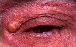
Molluscum contagiosum is a common viral skin infection caused by the molluscum contagiosum virus (MCV), a poxvirus. On the eyelids and periocular skin, it presents as small, flesh-colored, dome-shaped papules with a characteristic central umbilication, typically 2–5 mm in diameter. Lesions may be single or multiple. The condition is most common in children, immunocompromised individuals, and adults who acquire it through skin-to-skin contact.
Ocular significance: Periocular molluscum lesions located at or near the lid margin may shed viral particles onto the ocular surface, causing a chronic follicular conjunctivitis that is often misdiagnosed as allergic or viral conjunctivitis. The conjunctivitis typically will not resolve until the eyelid lesions are treated. Any patient with unexplained unilateral chronic follicular conjunctivitis should have the eyelid margins examined carefully for molluscum lesions.
Diagnosis is clinical based on the characteristic umbilicated appearance. Histology (if performed) shows eosinophilic intracytoplasmic inclusions — Henderson-Paterson bodies — in the epidermal keratinocytes.
Treatment options:
Lid margin lesions causing follicular conjunctivitis should be treated promptly. Immunocompromised patients may develop extensive or treatment-refractory molluscum requiring systemic management.
A chalazion (a blocked oil-gland lump) is the most common benign eyelid lump — see the dedicated Chalazion page. Note: a chalazion that recurs in the same spot should be biopsied to rule out sebaceous carcinoma.
Connect with a board-certified oculoplastic surgeon who specializes in benign eyelid lesions.
Search the Directory →