The Caruncle & Its Lesions
The lacrimal caruncle is the small pink nodule you can see at the very inner corner of each eye. It is normal, healthy tissue — but like any tissue it can develop growths. Most caruncular lesions are benign, yet the caruncle’s location at the medial canthus, right beside the tear-drainage system, means lesions here deserve a knowledgeable eye and careful removal when needed.
This is an in-depth companion to our main Eyelid Skin Tumors & Lesions guide. Caruncular growths are part of the broader family of eyelid lesions, and sit beside the tear-drainage (lacrimal) system.
Caruncle Anatomy
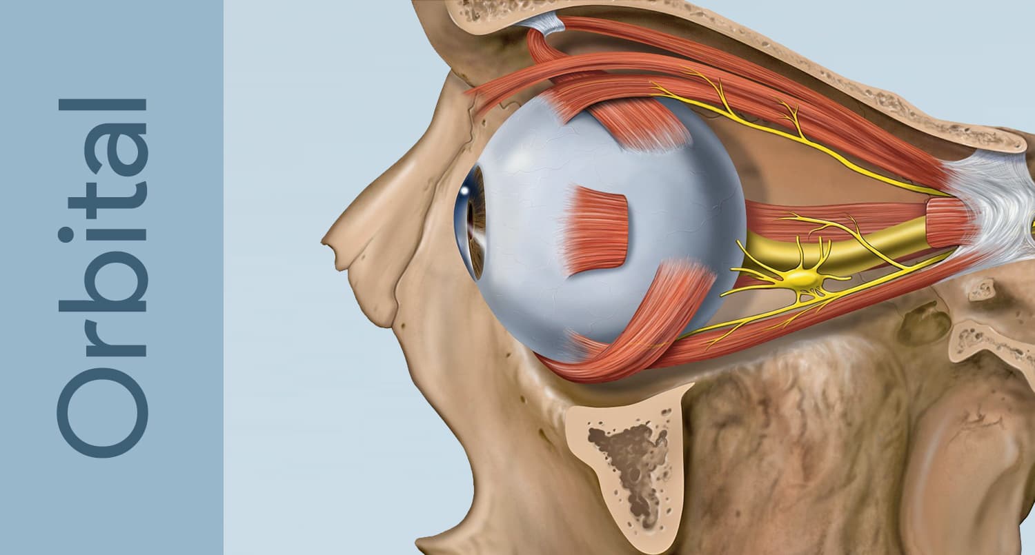
The caruncle sits in the inner corner of the eye, medial to the semilunar fold of conjunctiva. It is a small mound of modified skin and conjunctiva that contains fine hairs, sebaceous (oil) glands, sweat glands, and accessory lacrimal tissue. Just lateral and below it lie the puncta — the tiny openings that begin the tear-drainage pathway — which is why surgery in this region is planned to protect tear outflow.
Common Caruncular Lesions
- Papilloma — the most common caruncular growth; a soft, often frond-like benign lesion (see eyelid papilloma).
- Nevus (mole) — a pigmented benign lesion that may darken slightly over time, especially around puberty or pregnancy.
- Inclusion cyst — a fluid-filled benign cyst.
- Oncocytoma and other glandular lesions — uncommon benign growths of the caruncle’s glands.
- Malignant lesions — rare, but melanoma, sebaceous carcinoma, and other cancers can arise here, which is why atypical lesions are biopsied.
Warning Signs
Most caruncular lesions are harmless, but have one evaluated if you notice new or rapid growth, darkening or a change in color, surface irregularity or ulceration, bleeding, or a persistent feeling of fullness or irritation at the inner corner. Pigmented lesions that change are taken seriously because melanoma, although rare here, is possible.
Diagnosis
An oculoplastic surgeon examines the caruncle at the slit lamp, documents the lesion (often with photography to track change), and decides whether observation or biopsy is appropriate. Atypical, pigmented, or growing lesions are removed by excisional biopsy and sent for pathology to establish the diagnosis with certainty.
Treatment
Benign, stable lesions can simply be monitored. When removal is warranted — for symptoms, appearance, or to confirm the diagnosis — excisional biopsy is performed under local anesthesia, with care to preserve the adjacent puncta and canaliculi of the tear-drainage system. Larger or malignant lesions may require wider excision and reconstruction.
When to See an Oculoplastic Surgeon
See a specialist for any caruncular lesion that is new, growing, pigmented and changing, bleeding, or irritating. Because the caruncle abuts the tear-drainage system, removal is best handled by a surgeon trained in eyelid and lacrimal anatomy.
See an oculoplastic surgeon
Eyelid lesions sit millimeters from the eye, where removal and reconstruction demand specialist care. Find an ASOPRS-trained oculoplastic surgeon near you.