Ocular & Periocular Anatomy
Oculoplastic surgery is built on a detailed understanding of three interconnected anatomical regions — the eyelid, the lacrimal system, and the orbit. Select a region below for a comprehensive illustrated guide.
Interactive Anatomy Diagram
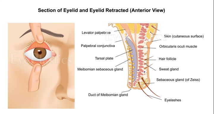
Section of Eyelid — Anterior View: skin, orbicularis, tarsal plate, Meibomian gland, palpebral conjunctiva, eyelashes
Eyelid Anatomy
Five tissue layers — skin, orbicularis, orbital septum, tarsus, and conjunctiva — working together to protect the eye and distribute the tear film.
Full anatomy guide →Lacrimal Anatomy
The complete tear pathway from production at the lacrimal gland through the puncta, canaliculi, lacrimal sac, and nasolacrimal duct into the nasal cavity.
Full anatomy guide →Orbital Anatomy
A bony pyramid roughly 40 mm deep housing the globe, six extraocular muscles, optic nerve, lacrimal gland, and orbital fat in intraconal and extraconal compartments.
Full anatomy guide →Eyelid Anatomy
Full guide →Key Structures
- Levator palpebrae superioris
- Müller's muscle
- Tarsal plate & Meibomian glands
- Canthal tendons (medial & lateral)
- Orbital fat compartments (upper & lower)
- Orbicularis oculi muscle
Lacrimal Anatomy
Full guide →Key Structures
- Lacrimal gland (superolateral orbit)
- Puncta (upper & lower lid margins)
- Upper & lower canaliculi
- Common canaliculus
- Lacrimal sac
- Nasolacrimal duct & valve of Hasner
Orbital Anatomy
Full guide →Key Structures
- Seven orbital bones (frontal, zygomatic, maxillary, sphenoid, ethmoid, lacrimal, palatine)
- Six extraocular muscles (four recti + two obliques)
- Intraconal & extraconal fat compartments
- Optic canal & superior orbital fissure
- Ophthalmic artery & superior ophthalmic vein
What is an oculoplastic surgeon?
Fellowship-trained specialists with expertise in all three anatomical regions — eyelid, orbit, and lacrimal system.